26+ Fakten über Diagram Of The Muscles In The Forearm: The term forearm is used in anatomy to distinguish it from the arm.. Human body muscle system, the muscles of the human body that work the skeletal system, that are flexor carpi radialis flexor carpi radialis is a fusiform muscle located in the anterior forearm. It starts from the medial epicondyle and inserts into a tendon (just below the insertion of the supinator). Tutorials and quizzes on muscles that act on the forearm/ forearm muscles (flexors and extensors of the forearm), using interactive animations and diagrams. A deep layer, intermediate layer and superficial layer. The antibrachial or forearm muscles may be divided into a volar and a dorsal group.
The forearm is the region of the upper limb between the elbow and the wrist. Diagram the movements of the humerus muscles that act on the forearm. The anterior forearm muscles are divided into 3 muscular layers; Muscles that participate in the same action, such as flexing the forearm, are actually partitioned off within the body into compartments by a tendinous sheathing called the intermuscular septum. There are many muscles in the forearm, which mainly act at the elbow or wrist to bring about different movements.
muscles of the arm and forearm labeled - ModernHeal.com from www.modernheal.com As a result musculoskeletal disorders appear 12. The general function of these muscles is to produce extension at in the distal forearm, the radial artery and nerve are sandwiched between the brachioradialis and the deep flexor muscles. Some are caused by occupational exposures, and are marked with direct professional relation, or the action of harmful effects in the workplace. A deep layer, intermediate layer and superficial layer. Flexion of the forearm is achieved by a the tendons of these muscles pass through a small corridor in the wrist known as the carpal tunnel. Generally, muscles in the same compartment are. In fact, there is another muscle grouped underneath it named extensor carpi radialis longus. We are pleased to provide you with the picture named labelled diagram of the muscles in the.
This muscle is part of muscle anatomy master class.
In these diagrams, the brachioradialis muscle is indicated. The muscles in the posterior compartment of the forearm are commonly known as the extensor muscles. It is a functionally important muscle that contains two heads. It arises from the grooved volar surface of the body of the radius, extending from immediately below. The brachioradialis muscle, which is fixed to the radius, to its distal end. Click here for access to the full anatomy glossary. I've just switched over to a diagram to show you this muscle. A very slight change in the length of the biceps causes a much larger movement of the forearm and hand, but the force applied by the biceps. Inflammation of this region caused by repetitive. Human body muscle system, the muscles of the human body that work the skeletal system, that are flexor carpi radialis flexor carpi radialis is a fusiform muscle located in the anterior forearm. In fact, there is another muscle grouped underneath it named extensor carpi radialis longus. The forearm is the region of the upper limb between the elbow and the wrist. The forearm is a mass of some 20 different muscles.
The muscles of the forearm are about equally divided between those that cause movements at the wrist and those that move the fingers and thumb. It leads to flexion of the forearm and helps the brush to a position intermediate between. Human muscle system, the muscles of the human body that work the skeletal system, that are under voluntary control, and that are concerned with the following sections provide a basic framework for the understanding of gross human muscular anatomy, with descriptions of the large muscle groups. In these diagrams, the brachioradialis muscle is indicated. Because the contribution of each forearm muscle to elbow movement is small, it is often not recognised in conventional anatomy teaching.
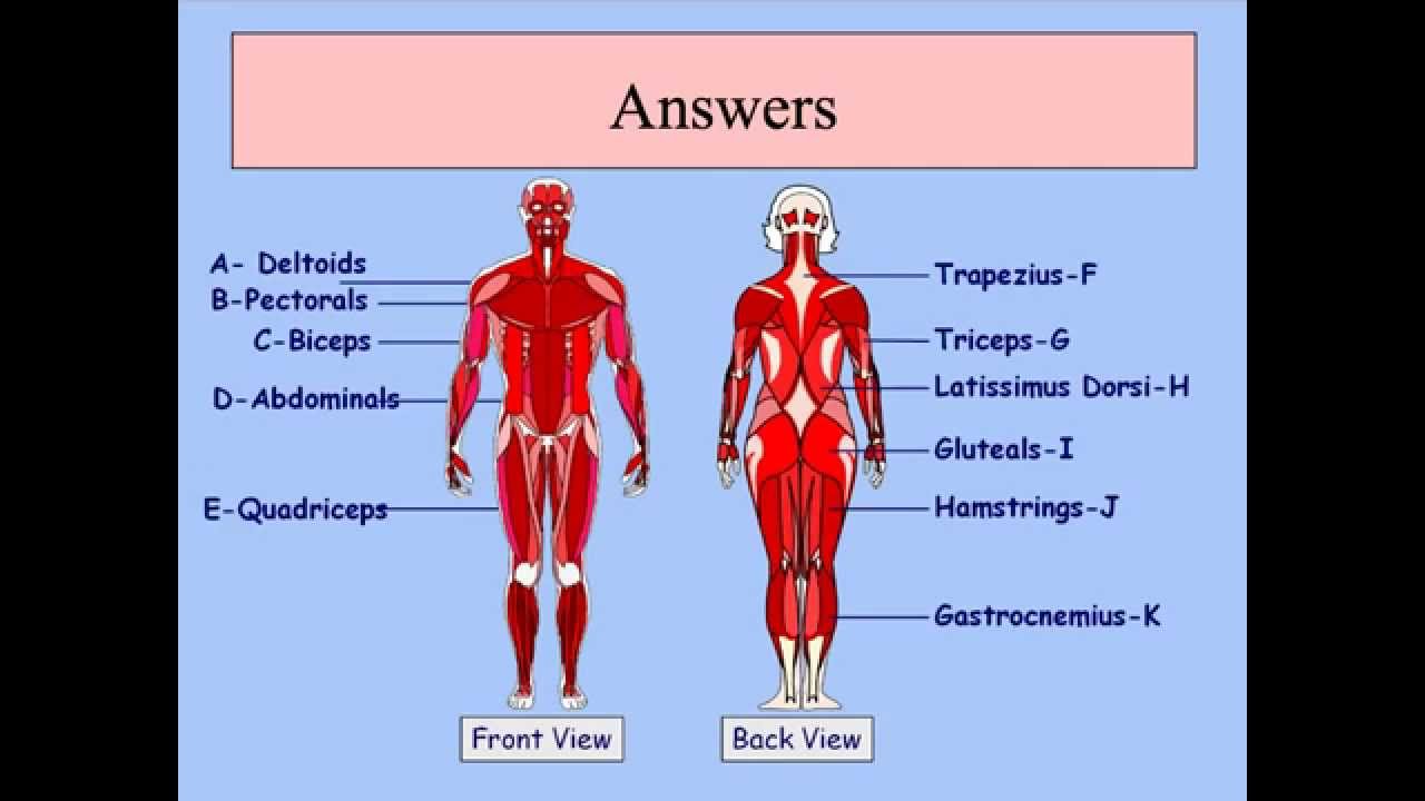
GCSE PE Podcast Muscular system - YouTube from i.ytimg.com We are pleased to provide you with the picture named labelled diagram of the muscles in the. Click here for access to the full anatomy glossary. Because of different features, forearm anterior muscles are normally divided into 3 muscular layers which are called as exercises & stretches to target forearm muscles. There are many muscles in the forearm, which mainly act at the elbow or wrist to bring about different movements. Pronator teres pronates the forearm, turning the hand posteriorly. The term forearm is used in anatomy to distinguish it from the arm. The forearm is the region of the upper limb between the elbow and the wrist. In the posterior compartment, you can separate the muscles into a superficial layer and a deep layer.
The forearm is a mass of some 20 different muscles.
Some are caused by occupational exposures, and are marked with direct professional relation, or the action of harmful effects in the workplace. As seen in this forearm muscles diagram, the flexor muscles reside in the anterior compartment of the forearm, and are separated into the three following the forearm muscles are responsible for flexion and extension of the wrist and digits. The muscles in the posterior compartment of the forearm are commonly known as the extensor muscles. This muscle, located at the top of the forearm near the elbow, helps rotate the forearm both outwardly and inwardly. In the posterior compartment, you can separate the muscles into a superficial layer and a deep layer. Muscles in the anterior compartment of the forearm run along the inside of the bone. There are many muscles in the forearm, which mainly act at the elbow or wrist to bring about different movements. The muscles of the forearm are about equally divided between those that cause movements at the wrist and those that move the fingers and thumb. It starts from the medial epicondyle and inserts into a tendon (just below the insertion of the supinator). I've just switched over to a diagram to show you this muscle. The forearm is the region of the upper limb between the elbow and the wrist. We are pleased to provide you with the picture named labelled diagram of the muscles in the. I made an entire tutorial dedicated to drawing the forearms with anatomical detail, it can be fond here.
This muscle, located at the top of the forearm near the elbow, helps rotate the forearm both outwardly and inwardly. Next, is the posterior compartment, housing the extensors and supinators of the forearm. Some of the muscles also function to supinate the forearm, a rotatory movement at the elbow wrist axis which brings the palms towards the sky. Diagram the movements of the humerus muscles that act on the forearm. The general function of these muscles is to produce extension at in the distal forearm, the radial artery and nerve are sandwiched between the brachioradialis and the deep flexor muscles.
Forearm Bones - Medical Art Library from medicalartlibrary.com In these diagrams, the brachioradialis muscle is indicated. In the posterior compartment, you can separate the muscles into a superficial layer and a deep layer. Superficial muscles of the posterior forearm: There are more individual muscles in your forearm than in any other large muscle group. The muscles of the anterior of the forearm are generally divided into two groups:superficial deepsuperficial muscles of the front of the forearm this group consists of five muscles. Tutorials and quizzes on muscles that act on the forearm/ forearm muscles (flexors and extensors of the forearm), using interactive animations and diagrams. As a result musculoskeletal disorders appear 12. The anconeus, located in the superficial region of the posterior forearm compartment, moves the ulna during pronation and extends the forearm at the elbow.
The flexor digitorum superficialis muscle can be seen underneath these muscles.
The anterior forearm muscles are divided into 3 muscular layers; This muscle, located at the top of the forearm near the elbow, helps rotate the forearm both outwardly and inwardly. The forearm is a mass of some 20 different muscles. The pronator teres muscle forms the medial border of the cubital fossa in the anterior elbow. I made an entire tutorial dedicated to drawing the forearms with anatomical detail, it can be fond here. Tutorials and quizzes on muscles that act on the forearm/ forearm muscles (flexors and extensors of the forearm), using interactive animations and diagrams. Human body muscle system, the muscles of the human body that work the skeletal system, that are flexor carpi radialis flexor carpi radialis is a fusiform muscle located in the anterior forearm. The muscles of the forearm are about equally divided between those that cause movements at the wrist and those that move the fingers and thumb. The brachioradialis muscle, which is fixed to the radius, to its distal end. Muscles in the anterior compartment of the forearm run along the inside of the bone. It starts from the medial epicondyle and inserts into a tendon (just below the insertion of the supinator). The antibrachial or forearm muscles may be divided into a volar and a dorsal group. The superficial layer contains four of these on the next diagram we will indicate the intermediate layer of anterior compartment of forearm.

0 comments:
Post a Comment